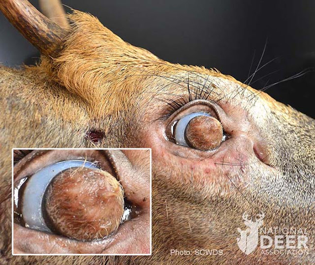
The report came in from Farragut, a suburb of Knoxville in east Tennessee, in late August 2020. “The individual stated the deer was circling, had visible bleeding, lacked awareness of the people around it, and had something on its eyes,” said wildlife biologist Sterling Daniels of TWRA.The deer was circling in a street, so local police and animal control officers were also alerted, responded first and dispatched the deer, saving the head at Sterling’s request, which he picked up later from a freezer. He shipped it to the Southeastern Cooperative Wildlife Disease Study unit (SCWDS) of the University of Georgia vet school... As you can see in these photos, instead of a normal cornea (the clear circle that includes the pupil and eye color), the buck had a disk of skin and dense hair completely covering the cornea of each eye. Microscopic examination at SCWDS determined the hairy growths were “corneal dermoids.”“Dermoids are a type of choristoma, which is defined as normal tissue in an abnormal location. Accordingly, dermoids are characterized by skinlike tissue occurring on the body in a location other than on the skin. Corneal dermoids, as in the case of this deer, often contain elements of normal skin, including hair follicles, sweat glands, collagen, and fat. The masses generally are benign (noninvasive) and are congenital, likely resulting from an embryonal developmental defect.”
More information at the website of the National Deer Association. Ocular dermoids have also been reported in dogs, and in humans - including on the cornea.

Limbal (corneal) dermoids are also found in humans!
ReplyDeletehttps://www.researchgate.net/figure/Grade-II-limbal-dermoids-involving-nearly-the-entire-depth-of-cornea-up-to-Descemets_fig2_236193456
I hadn't seen that. Thank you Amy. Added to the body of the post.
Delete